Userway menu

Pancreatic Cyst Surveillance

Pancreatic cysts are becoming increasingly common and the risk of them developing into pancreatic cancer is an important consideration.

Short Summary

  • Understanding pancreatic cysts is essential for proper management and surveillance.

  • Healthcare professionals can assess risk factors associated with pancreatic cancer to determine the most appropriate surveillance strategy for patients.

  • Patient perspectives should be taken into account to ensure patient-centered care tailored to individual needs and preferences.

At the Southern California Multi-Specialty Center we are experts at pancreatic cyst surveillance. If you suffer from pancreatic cysts, contact us for more information at 818-900-6480 for more information.

Let’s discuss all aspects related to these particular cysts. From types, methods for detection, their association with pancreatic cancer, and imaging techniques that come in handy when monitoring and surveying them.

We’ll also look into potential management strategies available to those patients diagnosed with a pancreatic cyst so they can understand what treatment options exist before making an informed decision on how best to move forward regarding their care.

Understanding Pancreatic Cysts

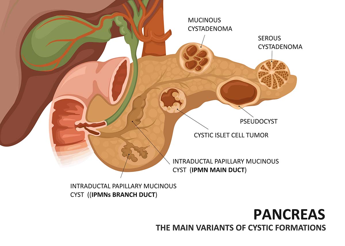
Illustration of the pancreas, showing the main variants of cystic formations

The incidence of pancreatic cyst disease is on the rise, with studies indicating a prevalence rate between 2% and 38%. This condition encompasses various types of fluid-filled sacs, ranging from benign cysts that do not lead to pancreatic cancer, to asymptomatic neoplastic pancreatic cysts that can precede high-grade dysplasia or even pancreatic malignancy.

Accurate diagnosis of the type of lesion is critical for determining effective treatment plans and follow-up strategies. The broad spectrum includes not only classic pancreatic cystic tumors but also other related types.

Types of Pancreatic Cysts

Pancreatic cysts are categorized into three types: pseudocysts, mucinous cystic neoplasms (MCNs), and cystic pancreatic neuroendocrine tumors (cNETs). Understanding the risk associated with each type is crucial for developing an appropriate management strategy for patients.

  • Pseudocysts do not pose a threat of malignancy. However, if a pseudocyst becomes infected, it can develop into a pancreatic abscess.
  • MCNs can potentially become malignant if certain characteristics are present, with a diameter of 3 cm or more being one of these significant indicators. Therefore, these cysts require appropriate surveillance.
  • Lastly, cNETs typically occur in adults between their 40s and 60s, accounting for around 15% of all pancreatic NETs. This makes them another potential candidate for ongoing monitoring.

Prevalence and Detection

The increasing prevalence of pancreatic cysts in recent years can be attributed to the advancements in imaging technology, which allows previously undetected cysts to be identified. This progress underscores the importance of regular monitoring, as early diagnosis and treatment can help prevent the development of pancreatic cancer.

Those who may require monitoring include individuals who have undergone surgery for the partial or complete removal of intraductal papillary mucinous neoplasms or distal pancreatic atrophy. Similarly, individuals diagnosed with incidental pancreatic cysts will likely need one or two consultations at the Southern California Multi-Specialty Center. This allows the risk factors to be evaluated before determining an appropriate monitoring and management plan for each unique case of cyst complications.

Increased awareness is crucial for potential patients concerning the necessary precautions due to the emergence and advancement identified through medical science. This is particularly important as these pancreatic cysts are appearing more frequently than before, often detected incidentally rather than during deliberate medical examinations.

Pancreatic Cancer Risk and Pancreatic Cyst Surveillance

A woman experiencing pelvic pain due to pelvic congestion syndrome

When dealing with pancreatic cysts, it is important to assess the risk factors associated with developing pancreatic cancer. By identifying these high-risk features, healthcare professionals can develop a plan for closely monitoring patients who have been diagnosed with this condition. Frequent surveillance and surgical intervention may be required if certain characteristics of the pancreatic cyst prove potentially dangerous in regards to possible instances of cancer down the line.

These particular risks must always be taken into account when assessing those who are affected by various forms of this type of cyclical condition. They can provide information which will help determine how best to approach treatment and reduce complications from arising due to its involvement in pancreas related diseases such as malignant tumors or carcinomas more generally.

High-Risk Features

Recognizing and assessing any high risk features associated with pancreatic cysts is essential for effectively determining a suitable management strategy. Such signs may involve an expansion of the main pancreatic duct (5 mm or more), having a size greater than 3 cm, presence of solid components/mural nodules, etc. In such cases, careful monitoring as well as surgical intervention might be required due to increased likelihoods of malignancy present within these elements. For each individual presenting with said issues, it’s important to keep track of their frequency in order to make sure appropriate treatments are made available promptly when needed.

Pancreatic Cyst Guidelines

Pancreatic cysts are a serious health concern, so it is important to recognize high risk features when evaluating patients. The American Gastroenterological Association (AGA) recommends that healthcare professionals consider more conservative options such as imaging surveillance rather than surgical resection for those with pancreatic cysts if deemed appropriate. This approach helps improve outcomes and provides the patient with an improved quality of life by tailoring care according to their individual preferences and risk factors.

Imaging Techniques for Pancreatic Cyst Surveillance

A CT scan of the pancreas to check for a pancreatic cyst
Pancreatic cyst surveillance necessitates the use of imaging techniques, such as CT scans, MRI with MRCP and endoscopic ultrasound (EUS), in order to observe any changes in size over time and detect potential signs of malignancy. These tools are critical for monitoring these particular types of growths accurately.

Computed Tomography (CT)

Pancreatic cyst surveillance is commonly conducted using computed tomography (CT) scans, an imaging technique which creates comprehensive images of the pancreas and surrounding structures. By emitting X-ray beams through the body, CT scan results reveal precise details about any existing pancreatic cysts. Such a procedure can be costly if not covered by insurance. It involves radiation exposure to the patient that could lead to potential harm in some cases.

Magnetic Resonance Imaging (MRI)

Due to its ability to detect small changes in tissue structure, MRI is an ideal choice for viewing pancreatic cysts and related features with great clarity. Compared to CT scans that use radiation, MRI offers a more appealing radiation-free option. It is not as easily accessible or cost effective and may be incompatible with certain medical conditions such as metal implants or pacemakers.

Endoscopic Ultrasound (EUS)

Endoscopic ultrasound (EUS) is a useful technique for evaluating pancreatic cysts, allowing the extraction of tissue samples through fine needle aspiration and giving detailed insights into their characteristics. This technique is most beneficial for cysts that are less than 2cm in size. The combination of endoscopy and ultrasound imaging provides extensive visuals within internal organs to aid in analysis.

Management Strategies for Pancreatic Cysts

A woman undergoing an ultrasound imaging to diagnose pelvic congestion syndrome
In regards to managing pancreatic cysts, there is a combination of observation, surgical intervention and modern diagnostic techniques used. Healthcare specialists will assess the type and nature of each individual’s cyst as well as any associated risks before deciding upon an appropriate monitoring plan that balances potential benefits with relevant risks related to pancreatic cyst surveillance.

Typically this begins by monitoring progress through regular imaging checks in order to detect changes or growths within the pancreas. While treatment may not always be necessary for such cases, observed care can help accurately evaluate their presence, which allows better-informed decisions around management of these areas depending on circumstances surrounding specific individuals who present with them.

Observation and Surveillance

Patients with pancreatic cysts that are not overly large or causing any discomfort and do not seem to be tumors should have observation through regular testing in order to establish if intervention is necessary.

It has been suggested that the first year requires check-ups every six months using either Magnetic Resonance Imaging (MRI) scans combined with Endoscopic Ultrasound (EUS), which then can drop down to annual tests for as long as no changes in the cyst take place. All of this serves as a way of keeping an eye on low risk cases involving such growths.

Surgical Intervention

When there is a high-risk pancreatic cyst causing significant symptoms, surgery might be needed. Guidelines suggest that if the cytology shows signs of malignancy or highly suggests dysplasia at a higher grade, then surgical removal should occur. Raised CA 19-9 levels in these types of cysts usually mean greater chances for cancer and may warrant additional tests as well as possible intervention through surgical intervention.

One must consider the risks along with potential advantages when looking into continued surveillance versus opting for surgery – it’s essential to weigh both options thoroughly before making any decision.

Emerging Diagnostic Modalities

Innovative diagnostics, such as next-generation sequencing and confocal laser endomicroscopy, are being studied to optimize the diagnosis and management of of pancreatic cysts. For example, by analyzing fluid collected from a pancreatic cyst it is possible to determine if the growth is benign or malignant. With this sort of advanced microscopy, it becomes feasible for practitioners to view molecular characteristics that aid in distinguishing between cancerous and non-cancerous lesions within a pancreas cyst.

Advances in technology like these paired with artificial intelligence have immense potential when considering enhancing patient outcomes through better diagnosis related issues around systency management.

Patient Perspectives on Pancreatic Cyst Surveillance

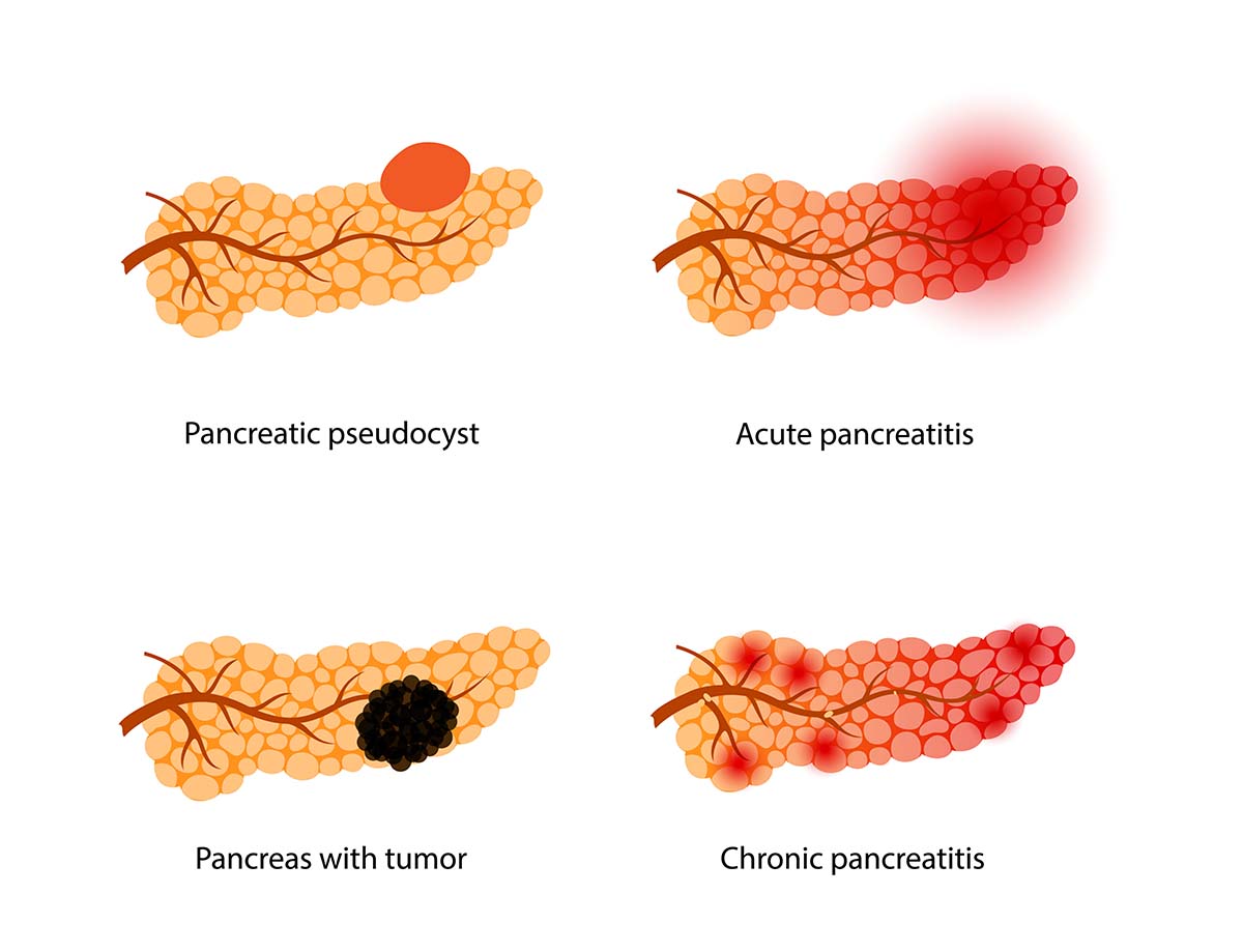
Illustrations of pancreas disease and cancer

It is important to take into account the patient’s viewpoint regarding pancreatic cyst oversight so as to deliver care that puts their needs first and evaluate advantages and risks of surveillance protocols. Patients might have worries concerning the size, texture or frequency of follow-up/intervention suggested by SCMSC.

To guarantee a decision is taken based on each individual’s requirements and preferences while still following recommendations from clinical guidelines committee and the Fukuoka guidelines and international consensus standards, health experts should include patients in this process.

Benefits and Risks

Patients should be educated on both the potential pros and cons of undergoing cyst surveillance for pancreatic cancer. Through this process, early discovery of tumors can take place along with tracking growth in existing cysts. There may also be risks such as false positives, radiation exposure or even unnecessary surgery related to it. SCMSC believes that providing patients with all the information available empowers them to make an informed choice about their care while giving them control over their own health.

Patient-Centered Care

The surveillance of pancreatic cysts requires a patient-oriented strategy to be effective. This involves examining risk factors, habits and any other elements that may have an influence on their choices by taking into account every individual’s needs, desires and priorities. By having such an approach at its core, SCMSC can have better results with greater patient satisfaction, as well as considering each person’s personal preferences when deciding upon what is suitable for them specifically regarding this condition.

Summary

Pancreatic cyst surveillance is essential for patient-centered care, providing insight for the growth of a cyst and its potential malignancy. Healthcare professionals must be well informed to accurately diagnose different types of pancreatic cysts with an array of imaging techniques suited to each case. Advances in diagnostic modalities could significantly enhance management practices by empowering both patients and practitioners alike.

With SCMSC’s personalized approaches that take into account individual needs, preferences and values, we can ensure quality medical assistance tailored specifically around one’s circumstances when it comes to managing pancreatic diseases. By staying up-to-date about what’s new concerning (pancreatic)cyst supervision methods and decisions related thereto. Those affected have control over their health status via educated judgments based upon available data relating thereto.

At the Southern California Multi-Specialty Center, the complexities associated with evaluation/surveillance involving these often difficult conditions can benefit even more people requiring such treatments!

Frequently Asked Questions

illustration of two types of abdominal aneurysm also known as stomach aneurysm

Do all pancreatic cysts need to be monitored?

It is essential to keep track of pancreatic cysts and carry out the right treatment depending on their size and texture detected by an MRI scan. Though rarely causing any issues, it is still important to observe them. Regular check-ups with a doctor are recommended in order for any possible changes to be spotted quickly and adequately treated if needed. It is also vital that one stays alert about potential symptoms suggesting there may be something wrong.

When do you stop surveillance pancreatic cysts?

It is suggested that close observation of pancreatic cysts stops if no changes in features have been detected over the period of 5 to 10 years. By following this advice, patients get optimal treatment and care. Surveillance should be terminated when there has not been any significant alteration within a 5 or 10 year time frame for these cysts.

How are pancreatic cysts monitored?

Pancreatic cysts are monitored using Magnetic Resonance Cholangiopancreatography (MRCP) imaging, which is considered the imaging test of choice. For cysts with higher-risk features, examination with Endoscopic Ultrasound (EUS) and Fine-Needle Aspiration (FNA) may also be recommended.

Followed by MRI testing every one or two years.

What are the different types of pancreatic cysts?

Pancreatic cysts come in three main types: pseudocyst, mucinous cystic neoplasms (MCNs), and neuroendocrine tumors (cNETs). These can range from small to large with varying levels of seriousness. They are symptomless but may cause abdominal pain, nausea, vomiting or jaundice. Treatment depends on the size and type as well as if it is malignant or benign. Both MCN’s and cNET’s pose risks for being cancerous, which need special consideration when creating a plan.

What are the high-risk features of pancreatic cysts?

The size of pancreatic cysts greater than 3 cm and the presence of a solid component or mural nodule, as well as dilation in the main pancreatic duct measuring 5 mm or more are all considered high risk features. This could suggest that there may be an existing instance of pancreatic cancer, which warrants Imaging evaluation or biopsy for confirmation. Any appearance exhibiting these characteristics should be treated with particular attention due to their potential danger.

Schedule an Evaluation

The SCMSC team in LA is always here, blending professional expertise with a touch of human understanding. To schedule an evaluation with Dr. Eghbalieh or Dr. Young at Southern California Multi-Specialty Center, call 818-900-6480.

Dr. Babak Bobby Eghbalieh is the colo rectal surgeon who treats colon cancer, bowel cancer and rectal cancer in Los Angeles

Babak (Bobby) Eghbalieh, MD, FACS

Dr. Stephanie Young of SCMSC

Stephanie Young, MD/MPH

Make an appointment at SCMSC

We look forward to welcoming you

Schedule an evaluation with Dr. Eghbalieh or Dr. Young at Southern California Multi-Specialty Center.

SCMSC front office staff in Los Angeles welcoming a new patient  for abdominal aortic aneurysm treatment