Carotid Evaluation Ultrasound
Carotid Ultrasounds for Los Angeles
What is a carotid ultrasound?
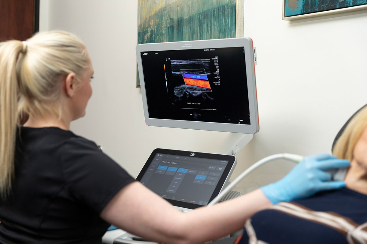
A carotid ultrasound is a painless, safe test that uses sound waves to make images of what your carotid arteries look like on the inside. By using the Doppler ultrasound technique, your healthcare provider can also see how well your blood travels through your two carotid arteries, which take blood to your brain.
A carotid ultrasound does not use radiation. It uses sound waves to make pictures of the inside of your carotid arteries. A computer takes the information from the transducer (the device your provider places on your skin) and uses it to make carotid ultrasound images on a monitor. The sonographer can record videos of the scans or save snapshots of them like photos.
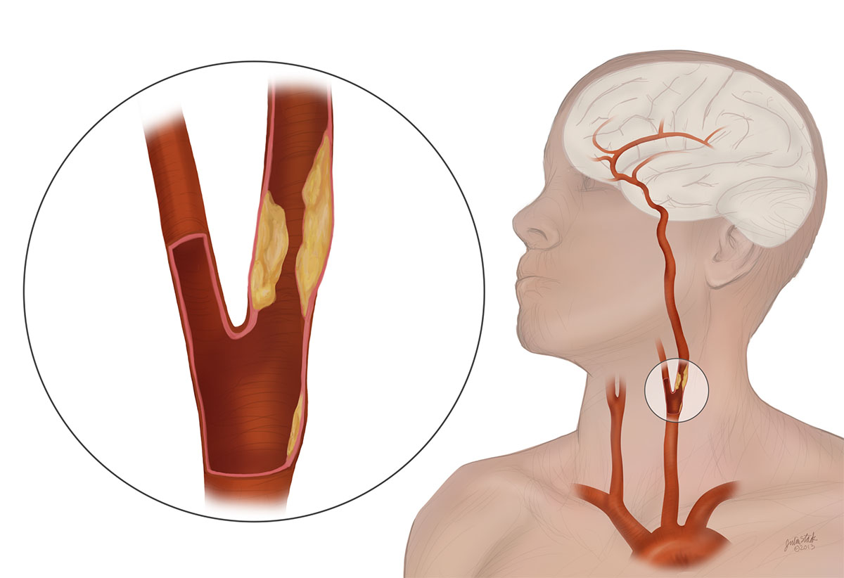
Normal Circulation
There are two arteries in your neck, the left and right carotid arteries. Each of these branch upward into the internal and external carotid arteries. These arteries are responsible for blood flow to your brain.
Any blockage of these arteries can result in brain damage or strokes.
Carotid Evaluation Ultrasound Q & A
What type of results do you get and what do the results mean?
If you get a normal result, your carotid arteries aren’t narrowed or blocked.
If your results aren’t normal, you may have atherosclerosis, a blood clot or some other problem that makes your artery too narrow and puts you at risk for a stroke.
If you have plaque buildup in one or both of your carotid arteries but the blockage is less than 50% (with stroke or TIA symptoms) or 60% (without stroke or TIA symptoms), your provider may tell you to improve your diet, exercise more and stop using tobacco products.
Also, your provider may prescribe medicine to:
- Dissolve blood clots (thrombolytics).
- Prevent blood clots (aspirin or clopidogrel).
- Lower your cholesterol level (statins).
- Bring down your blood pressure (antihypertensives).
If the buildup is more severe (at least 50% with stroke or TIA symptoms or 60% without stroke or TIA symptoms), they may recommend a surgical procedure.
A carotid ultrasound is generally accurate, but there can be times when it looks like there’s a blockage, but there isn’t one. The experience level of sonographers can vary, and ultrasounds do better at picking up severe problems than milder ones. Your provider may want you to have more imaging tests, such as cerebral, CT or magnetic resonance angiography. One reason you may need these other types of imaging is because of bone that blocks the ultrasound’s view of part of your carotid artery. The ultrasound can’t “see” through the bone.
Risk factors
Risk factors for strokes include smoking, salt intake, being overweight, not getting enough exercise, stress and taking birth control pills. Other risk factors include your age, gender, race and heredity.
What to expect during a carotid ultrasound
Your healthcare provider will put some clear gel on the sides of your neck. (You have one carotid artery on each side of your neck.) This gel might feel warm on your skin, but the gel makes it easier for sound waves to travel between your arteries and the transducer your provider uses.
For quality images during the test, it’s best not to move around.
While you’re lying on your back, your provider will press the transducer (smaller than a handheld price scanner at a store) against the skin on the sides of your neck. The sound waves coming out of and back into the transducer after hitting your arteries along the way will tell the computer what the image looks like. Your provider will keep moving the transducer over your skin until they have all the images they need.
You won’t hear the sound waves unless it’s a Doppler ultrasound. In that case, you may hear swishing sounds that change pitch. The procedure can take anywhere from 30-45 min.
What will I feel during the exam?
Don’t worry – since there are no needles or anything else inserted into your body, the test is completely painless and has no side effects. You will be asked to lie down on a stretcher. The technician will ask you several questions about why your physician ordered the exam. The recordings are received by running a small, smooth probe gently over various parts of your neck and jaw area. This will show if there is blockage in the large arteries of the neck.
At SCMSC we have an ultrasound tech onsite and can receive and review the images right away to save time. Other surgeons may refer you out to another office to receive the ultrasound and it may take several days for the results of the exam to be sent to your doctor.
Our Vascular & Endovascular Surgeons
It's important to remember not all physicians are trained in advanced vascular and endovascular surgery. It’s a good practice to get multiple opinions and do research on the surgery and the physician.
Make an appointment at SCMSC
We look forward to welcoming you
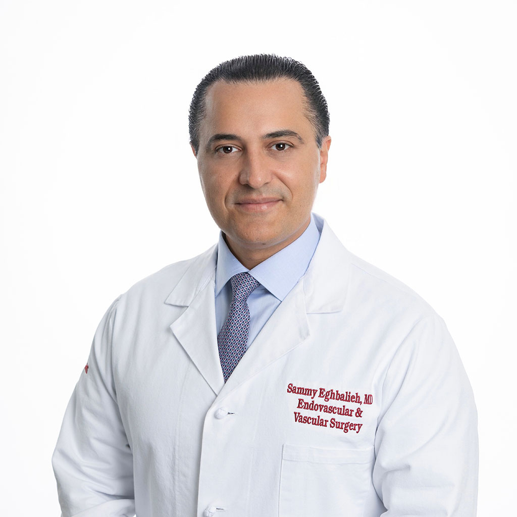
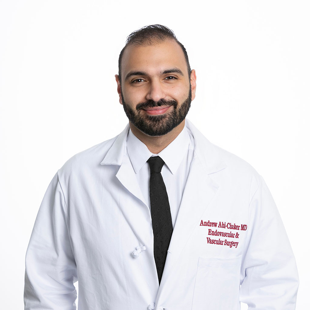
Schedule an evaluation with Dr. Eghbalieh or Dr. Abi-Chaker at Southern California Multi-Specialty Center.


